Digital devices for radiography have significantly changed the approach to diagnostics in dentistry. Previously, x-ray images in dentistry were created with the help of traditional films, which required not only a long process of obtaining images, but also significant exposure of the patient. Dentists had to manually process the films, which took a lot of time and also required additional costs for materials. In addition, at that time there was a problem with the accuracy of images, because it was not always possible to obtain sufficiently clear images for a correct diagnosis.
Technologies have changed today. Digital x-ray devices allow you to get images instantly and with minimal exposure. Modern digital sensors have replaced films, and data can be stored electronically, which greatly facilitates access to them and increases the efficiency of dentists' work. Now it is possible not only to obtain high-quality images, but also to integrate them into electronic medical systems for faster and more accurate diagnostics.
Target radiography is a method that allows you to get a picture of one or several teeth, focusing attention on a specific problem area. This method is extremely important for accurate determination of tooth disease and the subsequent appointment of individual treatment. Modern aiming pictures are as safe as possible, since the dosage of X-ray radiation is minimal.
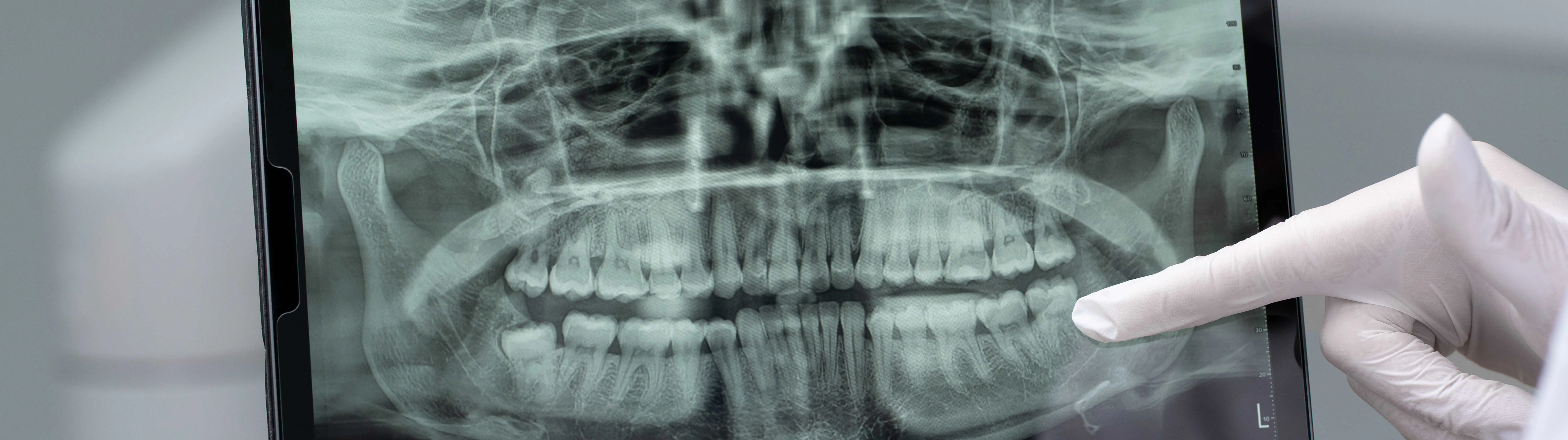
A panoramic image of the teeth (ortopantomogram) is one of the most popular radiography methods in dentistry, which allows you to get an image of the entire jaw. This method allows the dentist to assess the general condition of teeth, bone structures and soft tissues. For detailed study, the image is displayed on a computer monitor, where the dentist can enlarge the desired areas and examine them in more detail. Modern X-ray devices provide high-quality images that allow detecting even the smallest pathologies of the dentition in the early stages. This helps to start treatment in a timely manner and prevent the development of diseases.
Cone-beam computer tomography (CPCT) is the most modern method of radiography, which allows obtaining three-dimensional 3D images of the upper and lower jaw. The high accuracy of 3D images allows you to assess the course of treatment during dental implantation, aesthetic prosthetics, and bite correction. KPKT also makes it possible to accurately determine the structure and volume of bone tissue, which is critically important for successful implantation. Pictures allow you to clearly see the position of the mandibular nerve, which helps to avoid damage during operations. This method is also used to monitor the bite correction process, providing accurate prediction of treatment results.
Digital devices for radiography in dentistry have a number of significant advantages:
Reduced exposure. With the help of digital sensors, it is possible to obtain an image with a significantly lower dose of X-ray radiation.
Image processing speed. After the examination, the image appears on the screen almost instantly, which allows you to proceed to diagnostics.
High image quality. Digital technologies allow obtaining images with high resolution, which improves the accuracy of the diagnosis.
Ease of data storage and transfer. Digital images can be stored electronically, which facilitates archiving and access to the medical history.
Environmental friendliness. There is no need to use consumables, such as film, as well as chemicals for processing films. These factors reduce the negative impact on the environment.
Stationary devices for radiography are usually used in dental clinics with a large flow of patients. They provide high image quality and are used for a wide range of diagnostic tasks.
Advantages of stationary devices:
Reliability and stability of operation: these devices are designed for continuous operation, which makes them ideal for clinics with a large flow of patients.
Increased accuracy: thanks to the use of more complex and expensive components, stationary devices are able to provide higher accuracy of images.
A wide range of uses: they can be used for various types of examinations — from standard X-ray images to computer tomography or panoramic images.
Ease of maintenance: stationary devices usually require minimal maintenance.
However, stationary devices take up more space, and their installation requires special conditions, in particular to ensure safety from X-ray radiation.
Mobile x-ray machines are becoming more and more popular due to their convenience and the ability to conduct examinations directly in the offices. They are they are ideal for emergency situations or for clinics where high mobility and efficiency in conducting examinations are necessary.
Advantages of mobile X-ray machines:
Ease of transportation: this is an excellent solution for clinics with limited space.
Speed of installation: mobile devices can be quickly set up and start research without lengthy preparation.
Flexibility in use
Although mobile devices are convenient, they have limitations in the range of applications. They are mainly suitable for basic diagnostic studies and will be ineffective for complex examinations, for example, panoramic images or computer tomography. Therefore, stationary devices are used for more detailed or specific examinations.
Digital X-ray systems have significantly improved dentistry in diagnostics and treatment. The choice between stationary and mobile equipment depends on the specifics of the clinic and the needs of patients. It is important to carefully approach the choice of equipment to ensure the best quality of service and comfort for patients.